Antonie van Leeuwenhoek, a Dutch fabric merchant with no scientific training, ground his own lenses in his spare time and became, in 1674, the first person in human history to see a single-celled organism. He called the things he saw in his pond water "animalcules" — little animals — and reported them to the Royal Society in London, which initially did not believe him.
It is hard, from this remove, to imagine the conceptual shock of that observation. Until that moment, the world was thought to consist of things visible to the unaided eye and the things they were made of. Leeuwenhoek discovered that there was a third category: organisms too small to see at all, present in vast numbers in water, in soil, in our own bodies, an entire biosphere previously invisible to human awareness.
Everything we now know about microbiology, about disease, about cellular biology, about the actual mechanisms of life, descends from someone in seventeenth-century Delft grinding a small piece of glass.
What a microscope actually does
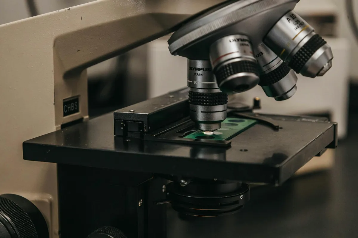
A microscope is, in the simplest description, an arrangement of lenses that produces an enlarged image of a small object. The basic optical principle is straightforward — a lens close to an object produces a magnified image, a second lens then magnifies that magnified image, and the eye perceives the final magnified result.
The technical difficulty is not in the basic principle. It is in the execution. Lenses must be ground to precise curvatures. Glass must be uniform and free of bubbles. Optical aberrations must be corrected through compensating lens designs. Light must be properly directed onto the specimen. Each of these problems took generations of patient work to solve.
By the late nineteenth century, the compound microscope had been refined to roughly the design used today: an objective lens close to the specimen producing the primary magnified image, an eyepiece magnifying that image, mechanical stages allowing precise positioning, condenser systems for proper illumination. The fundamental design has not changed substantially in over a century.
The diffraction limit
The classical microscope reaches a fundamental limit at about half the wavelength of visible light — roughly 200 nanometers. Below that scale, the wave nature of light prevents resolution regardless of how perfect the lenses are. This limit was understood by Ernst Abbe in 1873 and constrained microscopy for the next century.
The twentieth and twenty-first centuries developed several approaches to seeing below the diffraction limit:
Electron microscopy: using electrons rather than visible light, with much shorter effective wavelengths, allowing resolution into the nanometer range.
Scanning probe microscopy: physically scanning a probe across the specimen surface, producing images at near-atomic scale.
Super-resolution fluorescence microscopy: a set of clever techniques developed since the 1990s that work around the diffraction limit through fluorescent labeling and computational reconstruction.
Each of these enabled new scientific work that the classical microscope could not support.
The cultural significance
The microscope is not just a scientific instrument. It is a cultural object that fundamentally reshaped human understanding of biology, disease, materials, and the structure of matter itself. The germ theory of disease — that infection is caused by microscopic organisms rather than by miasmas or imbalances of humors — required the microscope to even formulate.
The development of public health, modern medicine, agriculture, food science, and materials engineering all depend on the cumulative work of looking closely at small things. The microscope is the foundational instrument of much of contemporary technical civilization.
This is sometimes obscured by familiarity. Most schoolchildren have used microscopes. The instrument has become ordinary. The achievement it represents — extending human perception by orders of magnitude — remains extraordinary.
The discipline of looking
What strikes me about microscopy as a practice, having watched experienced microscopists work, is that the instrument is the easy part. The hard part is the discipline of looking — knowing what to look for, what to ignore, what subtle features in a specimen indicate what underlying biological reality.
An experienced cytologist can examine a cell through a microscope and observe details that a novice would miss entirely. The instrument has not changed. The eye has not changed. What has changed is the trained perception — years of looking at thousands of specimens, learning what patterns mean what.
This is true of most observational sciences. The instrument extends human perception. The trained eye interprets what is perceived. Without the trained eye, the instrument shows interesting patterns whose meaning remains opaque.
The contemporary work
Microscopy continues to develop substantially. The past decade has seen advances in cryo-electron microscopy that have allowed determination of protein structures at near-atomic resolution. The 2017 Nobel Prize in Chemistry was awarded for these developments.
Live-cell imaging techniques have advanced to the point where individual molecules can be tracked moving through cells in real time. Multi-photon microscopy has extended optical microscopy into living tissue at depths previously inaccessible.
The instruments are not quite as cheap as the basic compound microscopes of the nineteenth century. Cryo-EM facilities cost millions of dollars to build and operate. Super-resolution systems require specialized expertise. But the cost is small compared to the scientific work they enable.
A closing observation
I find microscopy interesting partly because it is one of those fields where instrument and observation have developed in parallel for so long that they have become inseparable. You cannot do microscopy without microscopes. You also cannot do microscopy without the trained eye that can interpret what the microscope reveals.
The instrument extends what humans can perceive. The discipline interprets what is perceived. Together, they have produced most of what we know about the small-scale structure of the living and physical world.
The specimen sits on the stage. The lenses focus. The eye learns to see what is there.